Home
/ Abdominal Blood Vessels Labeled : 1 : Oxygenated blood is then returned to the left atrium of the heart by four pulmonary veins.
Abdominal Blood Vessels Labeled : 1 : Oxygenated blood is then returned to the left atrium of the heart by four pulmonary veins.
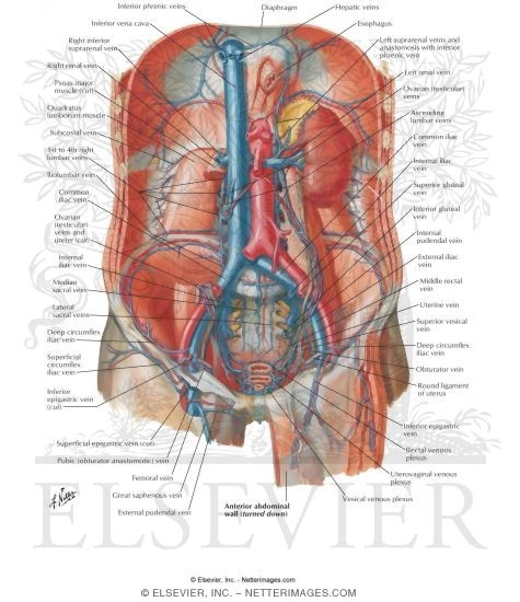
Abdominal Blood Vessels Labeled : 1 : Oxygenated blood is then returned to the left atrium of the heart by four pulmonary veins.. The abdominal aorta enters the abdomen through the diaphragm at the level of the twelfth thoracic vertebre and continues to just below the umbilical area, where it splits into the right and left common iliac arteries. Instant anatomy is a specialised web site for you to learn all about human anatomy of the body with diagrams, podcasts and revision questions Teachme anatomy part of the teachme series the medical information on this site is provided as an information resource only, and is not to be used or relied on for any diagnostic or treatment purposes. 3 the superficial vessels include the superficial epigastric and the superficial circumflex iliac vessels. In human anatomy, inferior epigastric artery refers to the artery that arises from the external iliac artery.it anastomoses with the superior epigastric artery.along its course, it is accompanied by a similarly named vein, the inferior epigastric vein.these epigastric vessels form the lateral border of hesselbach's triangle, which outlines the area through which direct inguinal hernias protrude.
The superior vena cava is the large vein that brings blood from the head and arms to the heart, and the inferior vena cava brings blood from the abdomen and legs into the heart. Label the abdominal blood vessels using the hints provided. Instant anatomy is a specialised web site for you to learn all about human anatomy of the body with diagrams, podcasts and revision questions Efferent branchial arteries injected with red latex. More posterior than the ivc until the umbilicus level where it lies more anterior than the ivc.
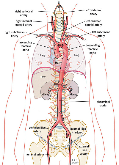
Veins Of Posterior Abdominal Wall Venous Drainage Of The Abdomen from www.netterimages.com That being said, all arterial blood delivered to this region comes via branches of the abdominal aorta, and all venous blood eventually finds its way back to. The abdominal aorta enters the abdomen through the diaphragm at the level of the twelfth thoracic vertebre and continues to just below the umbilical area, where it splits into the right and left common iliac arteries. If you continue browsing the site, you agree to the use of cookies on this website. Doppler studies of the abdominal vessels demand an understanding of normal and abnormal blood flow patterns. As the abdomen and pelvis contain the majority of internal organs, these regions need to be supplied by an extensive network of arteries and veins. Blood, the heart and the vessels that carry blood around the body together make up the cardiovascular system. Aortas or aortae 4) is the main blood vessel in the abdominal cavity that transmits oxygenated blood from the thoracic cavity to the organs within the abdomen and to the lower limbs. These vessels transport blood cells, nutrients, and oxygen to the tissues of the body.
Lateral view with the head to the right.
Instant anatomy is a specialised web site for you to learn all about human anatomy of the body with diagrams, podcasts and revision questions As the abdomen and pelvis contain the majority of internal organs, these regions need to be supplied by an extensive network of arteries and veins. Courses inferior through chest and enters abdomen through the diaphragm. Posterior cardinal sinus (blue) visible after lifting up the gi tract & gonads. We will include an analysis of the normal doppler waveforms of the abdominal vessels. (superficial epigastric visible at upper left.) the left femoral triangle. The portal venous system transports venous blood from the abdominal vasculature to the liver whilst the systemic venous system returns blood to the right atrium of the. These vessels are branches of the femoral artery and vein. Aortas or aortae 4) is the main blood vessel in the abdominal cavity that transmits oxygenated blood from the thoracic cavity to the organs within the abdomen and to the lower limbs. The superior vena cava is the large vein that brings blood from the head and arms to the heart, and the inferior vena cava brings blood from the abdomen and legs into the heart. The abdominal aorta enters the abdomen through the diaphragm at the level of the twelfth thoracic vertebre and continues to just below the umbilical area, where it splits into the right and left common iliac arteries. It specifically looks at the unpaired vessels of the abdominal aorta including the celiac trunk superior mesenteric and inferior mesenteric arteries as well as their branches. If you continue browsing the site, you agree to the use of cookies on this website.
Label the steps in the homeostatic response to high blood pressure. Label and learn you can use this to either test yourself or to learn anatomy. Students were given time t … Blood vessels the major vessels in the anterior abdominal wall can be divided into deep and superficial vessels (fig. More posterior than the ivc until the umbilicus level where it lies more anterior than the ivc.
The Aorta What Is It The Anatomy Images from my.clevelandclinic.org 3 the superficial vessels include the superficial epigastric and the superficial circumflex iliac vessels. This video series covers the blood vessels for anatomy and physiology ii students. It is an artery, meaning that it carries blood away from the heart. These vessels are branches of the femoral artery and vein. The abdominal aorta enters the abdomen through the diaphragm at the level of the twelfth thoracic vertebre and continues to just below the umbilical area, where it splits into the right and left common iliac arteries. (superficial epigastric vesseles labeled at center top.) details source femoral artery vein superficial epigastric vein identifiers latin arteria epigastrica superficialis mesh d019074 ta98 a12.2.16.011 ta2 4675 fma 20734 anatomical terminology [edit on. The superior vena cava is the large vein that brings blood from the head and arms to the heart, and the inferior vena cava brings blood from the abdomen and legs into the heart. The portal venous system transports venous blood from the abdominal vasculature to the liver whilst the systemic venous system returns blood to the right atrium of the.
The videos are done by dr.
As a medical student, i found anatomy pretty challenging. Blood vessels of the abdomen and pelvis. Aortas or aortae 4) is the main blood vessel in the abdominal cavity that transmits oxygenated blood from the thoracic cavity to the organs within the abdomen and to the lower limbs. The abdominal aorta enters the abdomen through the diaphragm at the level of the twelfth thoracic vertebre and continues to just below the umbilical area, where it splits into the right and left common iliac arteries. (superficial epigastric visible at upper left.) the left femoral triangle. Instant anatomy is a specialised web site for you to learn all about human anatomy of the body with diagrams, podcasts and revision questions Neurovasculature of the abdominal wall explore study unit superior epigastric artery: Anatomy of shoulder 12 photos of the anatomy of shoulder anatomy of nerves in shoulder, anatomy of posterior shoulder dislocation, anatomy of right shoulder, anatomy of shoulder labrum tear, anatomy of the shoulder games, human anatomy, anatomy of nerves in shoulder, anatomy of posterior shoulder dislocation, anatomy of right. The abdominal aorta is the largest blood vessel in the abdomen. Nodes drain to preaortic lymph nodes in root of primary arteries of gut (celiac nodes, superior and iferior mesenteric nodes) We will include an analysis of the normal doppler waveforms of the abdominal vessels. 3 the superficial vessels include the superficial epigastric and the superficial circumflex iliac vessels. If you continue browsing the site, you agree to the use of cookies on this website.
The superior vena cava is the large vein that brings blood from the head and arms to the heart, and the inferior vena cava brings blood from the abdomen and legs into the heart. It is an artery, meaning that it carries blood away from the heart. The aorta is the largest blood vessel in the body. Aortas or aortae 4) is the main blood vessel in the abdominal cavity that transmits oxygenated blood from the thoracic cavity to the organs within the abdomen and to the lower limbs. .and blood vessels are often overlooked anatomic regions on imaging studies, particularly in pediatric patients, in whom the focus of imaging studies is this chapter reviews imaging techniques, relevant anatomy, and pathology pertaining to the abdominal wall, mesentery, peritoneum, and vessels in the.
Blood Vessels Of Abdomen And Pelvis Anatomy Overview Kenhub from thumbor.kenhub.com As a medical student, i found anatomy pretty challenging. Blood vessels the major vessels in the anterior abdominal wall can be divided into deep and superficial vessels (fig. Courses inferior through chest and enters abdomen through the diaphragm. Want to learn more about it? More posterior than the ivc until the umbilicus level where it lies more anterior than the ivc. The videos are done by dr. Advertising on our site helps support our mission. It is an artery, meaning that it carries blood away from the heart.
Posterior cardinal sinus (blue) visible after lifting up the gi tract & gonads.
Anatomy of shoulder 12 photos of the anatomy of shoulder anatomy of nerves in shoulder, anatomy of posterior shoulder dislocation, anatomy of right shoulder, anatomy of shoulder labrum tear, anatomy of the shoulder games, human anatomy, anatomy of nerves in shoulder, anatomy of posterior shoulder dislocation, anatomy of right. Blood vessels of the abdomen and pelvis. Label the blood abdominal blood vessels labeled :. Introductory anatomy lab #8 slideshare uses cookies to improve functionality and performance, and to provide you with relevant advertising. Superficial epigastric artery scheme of the femoral artery. (superficial epigastric vesseles labeled at center top.) details source femoral artery vein superficial epigastric vein identifiers latin arteria epigastrica superficialis mesh d019074 ta98 a12.2.16.011 ta2 4675 fma 20734 anatomical terminology [edit on. Lateral view with the head to the right. It has a number of important relationships and branches, which very commonly appear in exam questions and anatomy spotters. (superficial epigastric visible at upper left.) the left femoral triangle. The portal venous system transports venous blood from the abdominal vasculature to the liver whilst the systemic venous system returns blood to the right atrium of the. Advertising on our site helps support our mission. Blood vessels the major vessels in the anterior abdominal wall can be divided into deep and superficial vessels (fig. Want to learn more about it?
Label and learn you can use this to either test yourself or to learn anatomy blood vessels labeled. Nodes drain to preaortic lymph nodes in root of primary arteries of gut (celiac nodes, superior and iferior mesenteric nodes)